Current case studies
Increasing exposure to radiofrequency electromagnetic fields (RF-EMF) has been the focus of public health concerns due to the rising daily use of technologies such as mobile phones. A mechanism of RF-EMF interaction with the human body is through heating. Current safety guidelines regarding RF-EMF exposures are based on the assumption that exposure to a RF-EMF at a whole-body average specific absorption rate (WBA-SAR) of 4 W/kg results in a 1 °C increase in core body temperature. Currently, there is insufficient knowledge to determine whether exposure to RF-EMF can result in other biological effects on the human body and thus requires investigation.
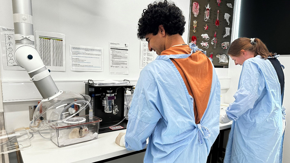
Recently, Deng and Rodney established a Bioelectromagnetics Laboratory in the Animal Facility at UOW. Our research starts with using improved methodology to accurately measure the thermal response of Sprague Dawley rats and C57BL/6 mice to RF-EMF. These studies have provided robust evidence showing that RF-EMF exposure at a high WBA-SAR (4-5 W/kg) increases core body temperature by 0.4-0.6 0C, which is less change than previously reported in these animals.
There have been concerns that RF-EMF exposure can negatively influence neurodegenerative diseases, like Alzheimer’s disease. On the other hand, there are studies suggesting RF-EMF exposure may alleviate the symptoms of such diseases. Using a mouse model of Alzheimer’s disease, we are currently investigating whether chronic exposure to RF-EMF at 5 W/kg WBA-SAR can influence the neuropathological hallmarks and cognitive symptoms of the disease.
Due to the increasing use of mobile phone in juveniles there is increasing concern that this population is more vulnerable to RF-EMF. While some studies have reported behavioral alterations following mobile phone use, we still do not have enough information regarding whether this is from the RF-EMF or other factors. Using a juvenile rat model, we are experimenting to see whether RF-EMF exposure at different WBA-SARs and exposure times during the childhood period can affect behavior and neurotransmission.

Research findings
- CHARACTERISING CORE BODY TEMPERATURE RESPONSE OF FREE-MOVING C57BL/6 MICE TO 1950 MHz WHOLE-BODY RADIOFREQUENCY-ELECTROMAGNETIC FIELDS
Emma Sylvester, Chao Deng, Robert McIntosh, Steve Iskra, John Frankland, Raymond McKenzie, Rodney J. Croft
- CHARACTERISATION OF THE CORE TEMPERAURE RESPONSE OF FREE-MOVING RATS TO 1.95 GHZ ELECTROMAGETIC FIELDS
Nathan Bala, Rodney J Croft, Robert L McIntosh, Steve Iskra, John V Frankland, Raymond J McKenzie, Chao Deng
Brain cancer poses an exceptional clinical challenge because of poor prognosis and side effects of treatment. It affects all age groups, including children, with many adults being in their most productive years. Many brain cancer survivors suffer from cognitive and somatic side effects of the treatment, with increased risks in children when cancer occurs before intellectual and physical development is completed. Glioblastoma is the most common and highly aggressive brain malignancy among all brain cancers. It requires potent treatments, including high-dose radiation therapy due to significant radio resistance. This highlights the need for new technologies and treatments for this disease. Producing more effective and synergistic chemo-radiation treatments that will improve survival and reduce morbidity would thus be clinically extremely valuable.
The effectiveness of novel nanoparticles and chemotherapy drugs was first assessed in vitro. After optimization of the in vitro parameters, the preclinical studies were developed in the Animal Facility at UOW.
Fisher rats were Computed Tomography scanned at the Monash Biomedical Imaging with iodine contrast to locate the brain tumor. The irradiation was performed using Synchrotron Microbeam Radiation therapy (MRT) at the ANSTO Australian Synchrotron. The radiation dose was delivered to nanoparticles localized in the brain tumor. Rats' long-term survival, investigated at the UOW Animal Facility, was the radiobiological endpoint.
Embracing change is needed, to impact brain cancer patient care. Our novel targeted nano-therapy using high dose rate Synchrotron Microbeam Radiation therapy and nanoparticles shows promise, increasing lethal damage specifically in tumor while sparing critical surrounding structures and reducing problematic unwanted side-effects.

Research findings
- Modulating Synchrotron Microbeam Radiation Therapy Doses for Preclinical Brain Cancer
Elette Engels, Jason R. Paino, Sarah E. Vogel, Michael Valceski, Abass Khochaiche, Nan Li, Jeremy A. Davis, Alice O’Keefe, Andrew Dipuglia, Matthew Cameron, Micah Barnes, Andrew W. Stevenson, Anatoly Rosenfeld, Michael Lerch, Stéphanie Corde & Moeava Tehei
- Toward personalized synchrotron microbeam radiation therapy | Scientific Reports
Elette Engels, Nan Li, Jeremy Davis, Jason Paino, Matthew Cameron, Andrew Dipuglia, Sarah Vogel, Michael Valceski, Abass Khochaiche, Alice O’Keefe, Micah Barnes, Ashley Cullen, Andrew Stevenson, Susanna Guatelli, Anatoly Rosenfeld, Michael Lerch, Stéphanie Corde & Moeava Tehei
- First extensive study of silver-doped lanthanum manganite nanoparticles for inducing selective chemotherapy and radio-toxicity enhancement
Abass Khochaiche, Matt Westlake, Alice O'Keefe, Elette Engels, Sarah Vogel, Michael Valcesi, N Li, KirrilyC. Rule, Josip Horvat, Konstantin Konstantinov, Anatoly Rosenfeld, Michael Lerch, Stéphanie Corde & Moeava Tehei
Graft-versus-host disease (GVHD) is an inflammatory disorder and major complication following donor stem cell transplantation in people with blood cancers and other blood disorders such as bone marrow failure. Use of donor stem cell transplantation is a curative therapy for blood cancers, as the donor immune cells can mediate a graft-versus-leukaemia (GVL) effect to combat any residual cancer in the patient. However, these donor immune cells (graft) can also mount an immune response attacking other tissues (liver, gut, skin and lung) in the recipient (host) leading to graft-versus-host disease (GVHD).
GVHD occurs up to 50% of patients following a donor stem cell transplant and can be fatal in up to 30%. Current therapies use broad range immunosuppression, leading to increased risk of cancer relapse and infection, therefore better therapies are required to treat GVHD.
We use preclinical mouse models that mimic the GVHD and GVL responses and allow us to test therapies to assess disease outcomes. These models are termed humanised mouse models, in which immunodeficient mice (no immune system) are engrafted with human immune cells and/or human leukaemia cells.
These preclinical humanised mouse models of GVHD and GVL developed in the Animal Facility at UOW allow us to examine the role of different human immune subsets, their engraftment and their role in tissue damage and anti-leukaemia immune responses. Furthermore, this allows us to test therapies to examine how to improve GVHD outcomes, while retaining the GVL effect.
These preclinical models allow us to further understand GVHD and potentially translate findings to improve the quality of life and outcomes for people with blood cancer and other blood disorders.
Research findings
- Post-transplant cyclophosphamide limits reactive donor T cells and delays the development of graft-versus-host disease in a humanised mouse model.
Sam Raj Adhikary, Peter Cuthbertson, Leigh Nicholson, Katrina M. Bird, Min Hu, Philip J O’Connell, Ronald Sluyter, Stephen I Alexander* and Debbie Watson* (*CO-SENIOR AUTHORS). - P2X7 receptor antagonism increases regulatory T cells and reduces clinical and histological graft-versus-host disease in a humanised mouse model.
Peter Cuthbertson, Nicholas J Geraghty, Sam R Adhikary, Sienna Casolin, Debbie Watson* and Ronald Sluyter* (*CO-SENIOR AUTHORS) - The P2X7 antagonist Brilliant Blue G reduces serum human IFN-γ in a humanised mouse model of graft-versus-host disease.
Nicholas Geraghty, Lisa Belfiore, Diane Ly, Sam Adhikary, Stephen Fuller, Winnie Varikatt, Martina Sanderson-Smith, Vanessa Sluyter, Stephen Alexander, Ronald Sluyter* and Debbie Watson* (*CO-SENIOR AUTHORS).
Motor neurone disease (MND) results in the selective death of upper and lower motor neurons in the motor cortex and spinal cord. MND patients develop progressive muscle weakness that eventually leads to death within 3-5 years following diagnosis and there are currently no effective treatments.
Mutations in the SOD1 gene are the second leading known cause of MND. These mutations subsequently lead to unstabilised formation of the SOD1 protein increasing the likelihood of it misfolding. SOD1 mouse models develop a progressive motor neuron disease that is highly similar to the human disease and provide a valuable insight into the basic biology of MND. Due to their ability to recapitulate the disease pathology of MND, SOD1 mouse models are commonly utilised to assess the preclinical efficacy of therapeutics in order for the safe transition into human clinical trials.
Ultimately, our current research at the UOW animal facility aims to guide the future design and testing of drugs that target SOD1 stabilisation to create effective treatment options for people diagnosed with MND.
Research findings
- CuATSM improves motor function and extends survival but is not tolerated at a high dose in SOD1G93A mice with a C57BL/6 background
- The PSX7 receptor antagonist JNJ-47965567 administered thrice weekly from disease onset does not alter progression of amyotrophic lateral sclerosis in SOD1G93A mice
- P2X7 antagonism using Brilliant Blue G reduces body weight loss and prolongs survival in female SOD1G93A amyotrophic lateral sclerosis mice


